Scientific rationale, state of the art beyond ARRIMAGE
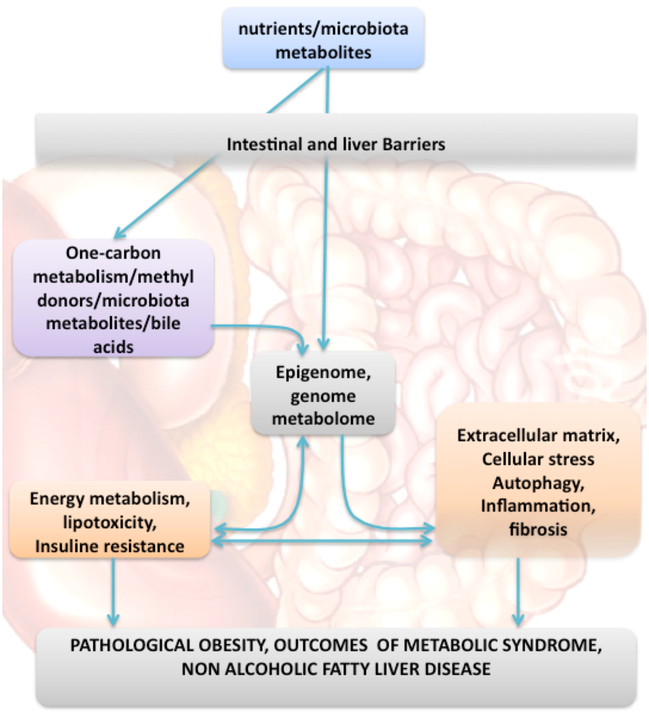
Our cutting-edge knowledge of pathological obesity and related diseases has progressed beyond whether we have good or bad habits, highlighting the importance of the complex interplay between genome, epigenome, microbiota, diet and related environmental risks. The pathogenesis of these diseases is complex and involves synergic pathomechanisms, including fetal programming, microbiota/metabolism crosstalk, disruption of the homeostasis of intestinal barrier and extracellular matrix, cell stress and dysregulation of genomic/epigenomic/host metabolism interplay.
The developmental origins of health and disease (DOHaD, fetal programming) consider that pregnancy and early post-natal life can have a long-term impact on later health and disease risk through gene-environment interactions and epigenomic mechanisms. Epidemiological studies have found imbalanced nutrition or methyl donor deficiency during pregnancy to be predictors of central obesity, insulin resistance and NASH in children (for review, Gueant et al., Trends Endocrinol Metab, 2013). Moreover, foetal programming and lipodystrophic central obesity are strong predictors of systemic manifestations of metabolic syndrome. It is noteworthy than inherited disorders of the one carbon metabolism (1-CM) and nutritional deficits in methyl donors during pregnancy are involved in similar epigenomic dysregulations associated with foetal programming and metabolic syndrome.
Gut microbiota is a master contributor to the risk of developing metabolic syndrome and NASH. In these diseases, high-fat diet, alterations of gastric acidity or intestinal motility produce changes in gut microbiota that influence energy harvesting from the diet and butyrate and methane production. Gut microbiota contributes to the improved metabolic phenotype after bariatric surgery with gastric bypass (Tremaroli et al, Nature 2012). The cross talk of host metabolism with gut microbial communities influences the serum level of metabolites such as uric acid level, PUFA and micronutrients, including folate, vitamin B12, vitamin A, vitamin C, vitamin E. For example, uric acid is closely associated with metabolic syndrome and NASH (Johnson R et al., Diabetes 2013). One third of uric acid is excreted into the gut where it is broken down by the bacterial 5- hydroxyisourate hydrolase. Uric acid causes mitochondrial stress that stimulates fat accumulation. The dysbiosis may increase the production of toxic bile acids. The ‘leaky gut’ produced by disruption of the homeostasis of the intestinal barrier plays a key role in pathological obesity and NAFLD. It contributes to low-grade visceral inflammation.
The extracellular matrix. Among all the components of extracellular matrix, elastin is a fascinating protein that could play a crucial role in metabolic syndrome and obesity, and that could lead to the discovery of innovative biomarkers and therapeutic targets (Wahart A. et al. FEBS J. 2019). It suffers from diverse post-translational modifications throughout life such as carbamylation and glycation products (Gorisse et al. PNAS 2016) as well as the release of elastin-derived peptides (EDPs) (Wahart et al. FEBS J 2019). The discovery of specific inhibitors of elastin receptor complex (ERC) will allow innovative pharmacological strategies. Moreover, EDPs and carbamylation and glycation –derived products could appear as robust cutting edge biomarkers.
The bioactive EDPs with ERC subunits (Bennasroune et al. Matrix Biol 2019), including peripheral Elastin Binding protein (EBP) associated to Protective Protein/Cathepsin A (PPCA) sub-unit. The third partner is the transmembrane Neuraminidase-1 (Neu-1), bearing a sialidase activity, and responsible for the signal transduction of the receptor. Interestingly, it is also involved in desialylation of Insulin Receptor (Blaise et al. Diabetes 2013), c-Met (Romier et al. Diabetes, 2018) or CD36 (Kawecki et al, Cell Mol Life Sci 2019), affecting their signalling activity. These modulations promote insulino-resistance along with obesity and NASH. Moreover, elastolysis could clearly be seen as a central factor of insulino-resistance as deficiency of elastases such as neutrophil elastase (NE) or cathepsin S (CathS) protect against fat accumulation and permit a better regulation of glucose homeostasis (Talukdar et al. Nat. Med 2012; Mansuy-Aubert et al Cell Metab 2013; Lafarge et al. Diabetologia, 2014).
Autophagy is a highly conserved homeostatic cellular mechanism that mediates the degradation of damaged organelles, protein aggregates, and invading pathogens via a lysosome-dependent pathway (Bonam et al. Nat Rev. Drug Discovery 2019). Autophagy exerts a primary role in coordinating cell metabolism and growth with environmentally induced stress. In the liver, it regulates hepatocyte functions and endothelial cells, macrophages and hepatic stellate cells. Autophagy modulation is recognized as a potential new therapeutic strategy to treat metabolic syndrome and NASH (Retnakumar & Muller 2019). A peptide called P140 that targets chaperone-mediated autophagy has shown promise in terms of safety and efficacy in patients with Lupus and model animals with autoimmune and non-autoimmune inflammatory diseases (Zimmer et al., ARD 2013; Macri et al., Autophagy 2015). Another peptide (LR12) that inhibits the triggering receptor expressed on myeloid cells-1 (TREM-1) restores impaired autophagy in experimental colitis (Kokten et al., J Crohns Colitis 2018). These two molecules have not been evaluated in metabolic syndrome and NASH, despite their influence on autophagy.
The crucial role of the epigenome can be evaluated by powerful methods analyzing the whole DNA methylome. An ARRIMAGE member discovered the association between the FTO gene and metabolic syndrome and the risk modulation by FTO methylation (Meyre D et al, Nature Genetics, 2009). Few epigenome wide association studies (EWAS) have been published so far for evaluating the association between DNA methylome, pathological obesity and NASH. An EWAS showed an association between increased BMI and methylation of HIF3A (Dick et al, The Lancet, 2014). A differential DNA methylation is observed in genes involved in liver fibrosis progression (Zeybel M et al, Epigenetics, 2015). DNA methylation produces remodeling signatures after bariatric surgery (Ahrens M et al, Cell Metab, 2013). Plasma DNA methylation is a potential biomarker for stratification of liver fibrosis and steatosis (Hardy T et al, Gut 2017; Nano J et al, Gastroenterology, 2017). We recently identified the methylation of plasma mSEPT9 as a novel circulating cell-free epigenetic biomarker to diagnose hepatocellular carcinoma in patients with NASH and cirrhosis (Oussalah et al, EBioMedicine, 2018).
Micronutrients and Nutritional methyl donors (folate, vitamin B12 and choline). The prevalence of micronutrient deficits appears higher in the obese subject, especially when associated with metabolic syndrome. The most frequent deficits concern vitamins B12, B9, B1, D, A and C (Sanchez A et al., Obes Surg 2016; Li Z et al., Clin Nutr. 2018). The two main dietary methyl donors, vitamin B12 and folate are directly involved in the synthesis of S-adenosylmethionine, the universal methyl donor for DNA and protein methylations. Thus, dysregulation of the one carbon metabolism (1-CM) can alter the epigenomic regulation of gene expression (Gueant et al, Trends Metab Endocrinol, 2013). Epidemiological studies and inherited diseases revealed an association between 1-CM and NASH. The methyl donor deficiency (MDD) during pregnancy and lactation yields liver steatosis in rat pups through altered methylation of PGC1-a and dysregulation of PPARs and HNF-4α (Pooya S et al, J Hepatol 2012) and stress of endoplasmic reticulum (ER) through down-regulation of sirtuin 1 (SIRT1) in MDD (Garcia MM, J pathol, 2011; Melhem et al, Gut 2016, Battaglia et al, Nucleic Acid Research, 2018). It is noticeable that decreased SIRT1 is a common hallmark of overnutrition and MDD.